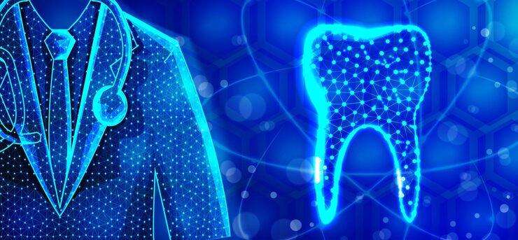
Light physics advances have revolutionized modern dentistry by enabling early detection of tooth decay and with unprecedented accuracy through non-invasive methods. Because of cutting-edge scientific understanding of dental tissue scattering and light absorption, cutting-edge optical caries detection technologies now outperform traditional radiography. Clinicians now have at their disposal safer, radiation-free diagnostic instruments that provide greater comfort and assurance to patients in diagnosis. This blog explores how light physics advances in non-invasive diagnosis are made operational, improving the detection of proximal caries and cracked tooth syndrome at earlier stages. It is central to an understanding of the behavior of light in interaction with dental hard tissues in developing such sensitive instruments, like Diagnoscan, that utilize new optical concepts in the identification of minute decay before it becomes irreversible.
Advances in Light Transmission and Scattering in Dental Tissues
Understanding dental tissue scattering is vital to the success of optical caries detection appliances. Healthy enamel permits light to pass through efficiently, but affected areas increase scattering due to the disrupted microstructure of the carious tissue. Light physics advances indicate that some wavelengths in the near-infrared spectrum are able to penetrate further into dental enamel with enhanced contrast due to differences in scattering from healthy and affected tissue. Measurement coefficient scattering experiments have shown that dental tissue scattering is substantially different between demineralized and healthy enamel (Fried et al., J Biomed Optics, 2022).
These scientific discoveries enable devices to be built that very tightly control light intensity and wavelength to optimize penetration and contrast, enhancing sensitivity in non-invasive diagnostics. Additional understanding of light physics advances in tissue interaction is a critical cornerstone to developing compact precise instruments like Diagnoscan, designed to highlight carious lesions by exploiting these scattering properties with eerie precision.
Optical Physics and Caries Detection
The optical caries detection mechanisms are based on differences in the amount of scattered and absorbed light by healthy and carious dental tissue. Attenuation of light occurs as the damaged areas scatter and absorb greater amounts of light, producing distinguishable shadows upon transillumination. Based on the developments in light physics, research has shown that such light signals provide high contrast images that detect early caries undetected by standard X-rays (Amaechi et al., Lasers Med Sci, 2023).
With sophisticated non-invasive diagnostic methods based on these principles, clinicians detect subsurface decay and cracks more reliably. Use of highly concentrated, focused light and low-noise photodetection ensures subtle differences in dental tissue scattering are translated into unequivocal diagnostic conclusions. This highly efficient optical interaction is the basis for the enhanced sensitivity of devices such as Diagnoscan, allowing dentists to detect and treat problems earlier and improve prognosis and patient comfort.
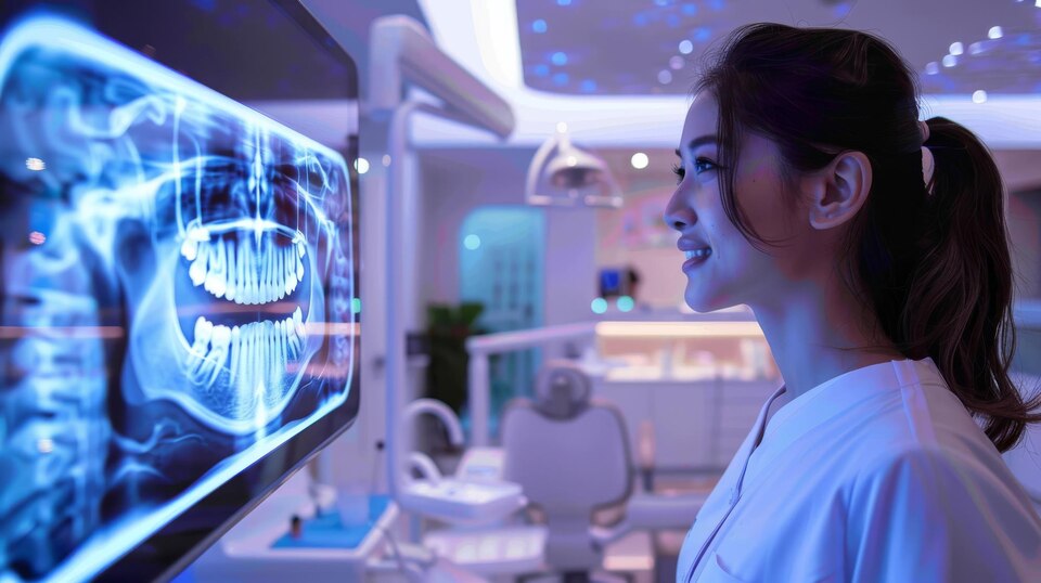
Non-Invasive Diagnostic Technologies
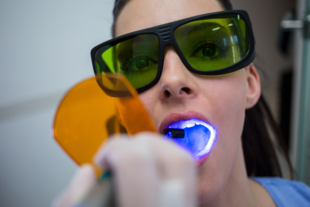
Non-invasive diagnostics now constitute an essential part of dentistry, reducing the application of exposure to radiation but increasing patient comfort and convenience. The method applies light physics advances to create high-intensity, well-controlled light beams, which penetrate dentin and enamel with minimal discomfort. They benefit from variations in dental tissue scattering to detect initial decay that are not detectable with regular X-rays.
Modern optical equipment has embedded microprocessors within it that are able to instantly analyze scattering patterns, thereby enabling optical caries detection in real time with precision. Internet of Things (IoT) connectivity takes it further by enabling remote sharing and tele-diagnosing, an advancement toward reaching more with dental care, especially in remote areas (Patel et al., Telemed J Health, 2021). The synergy of scientific understanding of light physics advances and innovative data transfer technologies places non-invasive diagnostics in a better position than radiography, which offers broader dissemination of rapid, trustworthy dental health assessment.
Scientific Validation of Light-Based Detection
There is uniform scientific evidence from numerous studies justifying optical caries detection methods developed through light physics advances. Such techniques are more specific and sensitive than traditional radiography, particularly in detecting caries and cracks that are hard to identify (J Clin Periodontol, 2024). Clinical trials have invariably demonstrated that instruments guided by dental tissue scattering and absorption understanding produce fewer false negatives and improved treatment outcomes.
Such widespread acceptance creates the case for bringing non-invasive diagnostics into general practice to augment and even supplant X-rays. The resultant decrease in radiation exposure and increased comfort for patients has significant public health implications, so optical caries detection is a scientific breakthrough of utmost importance.
Future Trends in Light Physics and Dental Diagnostics
The future of optical caries detection is characterized by ongoing research that integrates trends in light physics advances with emerging breakthroughs in artificial intelligence (AI), nanotechnology, and bio-optics. Emerging studies focus on the optimization of light sources and detection wavelengths to ensure maximum sensitivity to even the first changes in dental tissue structure. AI-driven algorithms integrated into optical diagnostics promise to present improved interpretation of complex dental tissue scattering patterns for increased diagnostic confidence and reduced human error.
Technological advancements in nanotechnology allow greater miniaturization of sensors and greater photodetector efficiency, enabling portable, low-cost, easy-to-use non-invasive diagnostics (Smith et al., Nanomedicine, 2023). In addition, imaging innovations in multi-modal imaging, such as transillumination with fluorescence or reflectance spectroscopy, offer more detailed information concerning tooth health. These light physics advances pave the way for the next generation of diagnostic systems that will allow clinicians worldwide to detect decay earlier, leading to enhanced, preventive dentistry with reduced invasive treatments.
Conclusion
Scientific research into light physics advances fundamentally transforms dental diagnostics by exploiting the intrinsic optical properties of dental tissues. Through detailed understanding of dental tissue scattering and absorption, clinicians can now leverage highly sensitive, non-invasive technologies for early tooth decay detection, surpassing the limitations of traditional radiographs. The integration of these scientific principles into practical devices ensures safer, radiation-free diagnostics that enhance patient comfort and accessibility. Tools such as Diagnoscan manifest this scientific progress, embodying advanced optical physics within compact, efficient diagnostic systems.
As AI, nanotechnology, and IoT connectivity further integrate with optical detection, the future promises increasing precision and ease of use. Ultimately, optical caries detection guided by ongoing light physics advances will continue to revolutionize dental care, facilitating early, minimally invasive interventions and improving health outcomes for patients worldwide.