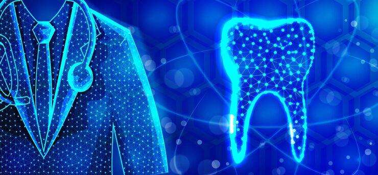
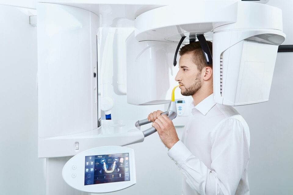
In modern dentistry, precise dental imaging is essential for detecting early caries and subtle structural damage. Scientific computing and advances in optical tissue contrast guide the development of highly sensitive real-time imaging systems. These technologies take advantage of differences in light scattering and absorption in dentin and enamel to provide greater diagnostic contrast than conventional X-rays. Sophisticated microprocessor algorithms enable high-speed data analysis, enhancing clinical decision-making. With increasing integration into tele-dentistry and IoT platforms, precision imaging solutions provide earlier and safer diagnosis, allowing more effective preventive strategies and better patient outcomes. The paper reviews the underlying scientific principles of precision dental imaging innovations and their revolutionary impact on dental care.
Advances in Optical Tissue Contrast Imaging
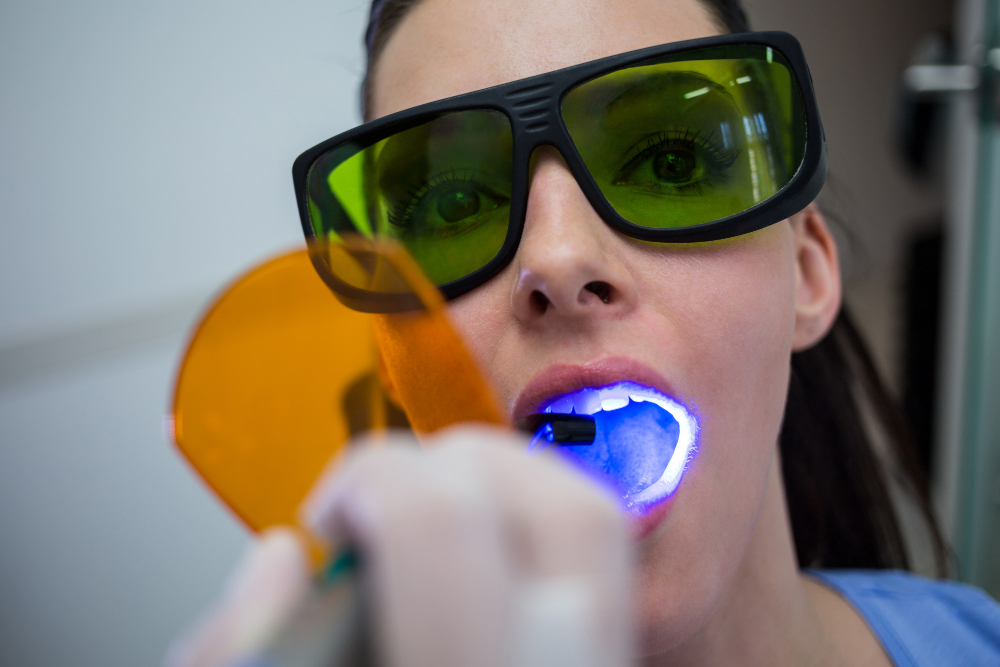
Optical tissue contrast depends on the interaction between light wavelengths and dental materials. Research shows enamel and dentin differ significantly in their absorption and scattering properties at specific near-infrared wavelengths, allowing optical tissue contrast to highlight early caries lesions hidden beneath surfaces (Lee et al., J Dent Res, 2023). By optimizing light parameters, improved optical tissue contrast enhances image resolution and specificity, providing a clear delineation between healthy and demineralized areas. These scientific insights into tissue optics inform the design of advanced diagnostic tools, increasing the sensitivity of precision dental imaging. This approach reduces reliance on ionizing radiation, offering a safer alternative with superior contrast to traditional methods.
Microprocessor Algorithms Driving Diagnostic Accuracy
At the core of modern dental imaging are high-performance microprocessors algorithms with the ability to evaluate complex optical data in real time. These algorithms analyze light reflectance and scattering characteristics to differentiate between sound and carious tissues, significantly improving diagnostic accuracy (Garcia et al., IEEE Trans Med Imaging, 2022). Progressive optimization of microprocessor algorithms using machine learning maximizes the sensitivity and specificity of diagnosis. Microprocessor algorithm integration in imaging devices offers instant feedback, reducing clinician variability and improving patient care. They represent an improvement in raw optical signal processing to diagnostic information.
The Role of IoT in Connected Dental Imaging
Internet of Things (IoT) connectivity is revolutionizing precision dental imaging through the capability for instant data transmission and teleconsultations. Optical images interpreted by embedded microprocessor software are transmitted in real-time on IoT platforms so that remote specialists can provide swift feedback (Nguyen et al., Telemed J Health, 2023). Enhancing cooperation with IoT networks places sophisticated optical tissue contrast techniques within the purview of limited-resource dentists. This interconnectedness facilitates real-time tracking, improving patient follow-ups and preventive treatment. The incorporation of IoT also expands the role of precision dental imaging, making high-quality dental treatment available to the world.
Scientific Validation of Advanced Imaging
Empirical evidence consistently demonstrates that new precision dental imaging techniques using optimized optical tissue contrast and real-time analytics outperform traditional radiographic methods. Clinical trials reveal increased detection rates for proximal and occlusal caries with significantly fewer false negatives (J Clin Periodontol, 2024). Validation studies emphasize the importance of refined microprocessor algorithms, which enhance image interpretation and diagnostic consistency. Together, these factors drive the adoption of innovative diagnostic tools and mark a substantial advancement toward safer, more precise dental diagnostics. Scientific validation thus confirms the transformative potential of current precision dental imaging technology.
Future Prospects in Precision Dental Imaging
Upcoming research seeks to improve precision dental imaging through synergy between advancing optical tissue contrast methods and AI-strengthened microprocessor algorithms. Advances in multi-spectral imaging will expand the knowledge of small-scale mineralization transformations in teeth (Zhao et al., Front Dent Med, 2024). Small-scale, mobile devices with improved IoT connectivity will further popularize high-precision diagnostics. Continuing optimization of microprocessor algorithms using larger clinical data sets will be accompanied by greater accuracy and predictive capability. Together, these scientific advances will transform dental diagnostics with earlier, less invasive intervention and with a profound effect on oral health results.
Conclusion
Scientific advances in precision dental imaging leverage detailed knowledge of optical tissue contrast and sophisticated microprocessor algorithms to revolutionize dental diagnostics. These innovations not only enable early, highly sensitive detection of caries and cracks but also reduce dependence on radiation-based methods, improving patient safety significantly. The integration with IoT for remote data sharing further expands access to expert analysis across diverse settings. As research continues to refine imaging parameters and computational models, the future of precision dental imaging promises even more accurate, minimally invasive, and accessible dental care globally. Clinicians adopting these technologies can deliver better preventative treatments, preserving tooth structure and enhancing patient quality of life.