Cracked tooth syndrome is traditionally hard to diagnose, particularly in its initial stages. Symptoms masquerade as other dental problems, meanwhile conventional imaging methods such as X-rays fail to detect microfractures given their low resolution and two-dimensional format. Advances in optical engineering, however, present a hopeful exception. Systems based on high-intensity lighting, such as Diagnoscan, enable the detection of minute cracks and stress lines in enamel and dentin layers. This blog takes an in-depth look at the science of dental microfracture detection, examining how advances in light-based imaging are transforming clinical decision-making. With research-supported protocols and enhanced visualization capabilities, optical diagnostics are fast becoming the go-to for dentists seeking accuracy.
Cracked Tooth Syndrome
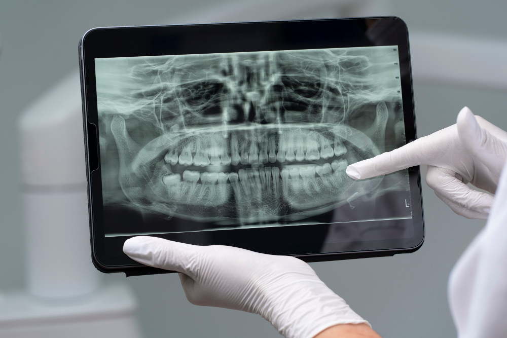
Cracked tooth syndrome is an incomplete tooth fracture, typically involving enamel and, at times, dentin. Clinically, it may manifest as diffuse pain on biting, temperature sensitivity, or referred pain, symptoms that make diagnosis challenging. Radiographs routinely fail to detect such cracks since they are mesiodistally oriented and too thin to deflect sufficient X-ray radiation. That’s where dental microfracture detection by light technology comes into its own. Optical techniques light up the internal structure of the tooth, accentuating thin fracture lines via differential light scattering. Current research (International Endodontic Journal, 2023) validates that optical detection raises diagnostic sensitivity in vertical and oblique fractures by over 30% in comparison to radiography.
Optical Scattering and Crack Visualization
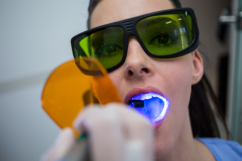
One of the fundamental principles facilitating successful dental microfracture detection is light scattering. Light travels with little interference when it enters a healthy tooth. A microcrack, however, interferes with this pathway, producing localized scattering and absorption. This abnormality is detectable using high-lux transillumination devices such as Diagnoscan, especially at wavelengths of 780-950 nm, which are ideal for internal dental imaging. Cracks are displayed as thin, darkened lines in illuminated images. This cannot be achieved through traditional X-rays, which possess poor spatial resolution. Developments in light propagation models have enabled the design of advanced dental diagnostic devices targeting specific wavelengths to maximize contrast in cracked areas.
The Role of Light Physics in Fracture Profiling
Fracture propagation in dental tissues is more than visualization, it is about stress distribution analysis and crack behavior. Optical methods like polarization-sensitive imaging and high-dynamic-range light mapping are being integrated into advanced dental diagnostic devices. These allow fracture width, depth, and direction profiling. Research in Applied Optics (2022) showed that polarizing filters with near-infrared transillumination could precisely measure crack depth up to 1.5 mm in enamel. Such detail is important for deciding the restorative approach. Diagnoscan-like devices are progressively incorporating such capabilities to facilitate total diagnostic mapping.
Lasers vs. Optical Scanners
Laser fluorescence devices have also been researched for dental microfracture detection, yet their application in the detection of cracks is limited by their reliance on bacterial activity for producing a signal. Optical transillumination is not dependent on bacteria being present but only on physical discontinuities in the tooth. Optical scanners are consequently more reliable in the detection of incipient cracks. Advanced diagnostic devices used in dentistry based on the use of IoT functions and micro-imaging processors, such as Diagnoscan, even allow storing images and longitudinal examination of them, so they are ideal for monitoring progressive development of crack growth. Laser equipment is excellent to detect active caries but lags behind in detecting purely mechanical failures.
Clinical Implications and Preventive Measures
The potential to identify microfractures early on has direct clinical relevance. If left undiagnosed, microcracks may propagate, ultimately causing tooth splitting, loss of structure, and more invasive treatments. Optical dental microfracture detection systems enable practitioners to intervene earlier with conservative restorations or occlusal adjustments. In preventive dentistry, these systems aid in risk assessment and patient education, establishing a more robust trust model. In addition, when coupled with digital records, advanced dental diagnostic devices make second opinions and case reviews easier. Technologies such as Diagnoscan are at the forefront of this evolution, moving dental care toward proactive management.
Conclusion
Optical imaging is not just a tech trend, it’s a dental diagnostic paradigm shift. For diagnoses as elusive as cracked tooth syndrome, trusting the science of light-scattering through advanced dental diagnostic devices is both evidence-based and practice-enhancing. As technology such as Diagnoscan becomes more prevalent in clinic routines, the benchmark for early, accurate, and radiation-free diagnosis is considerably raised. The science of dental microfracture detection not only enhances patient care but redefines the practice of conservative dentistry. For forward-thinking clinicians, investing in optical innovation is no longer a choice, it’s a necessity.